TearScienceTM LipiViewTM II Ocular Surface Interferometer
TearScienceTM LipiViewTM II Ocular Surface Interferometer
Leading innovation in lipid layer thickness measurement and meibomian gland imaging.

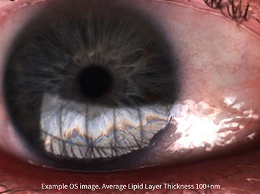
TearScience™ LipiView™ II Ocular Surface Interferometer with Dynamic Meibomian Imaging™ (DMI) measures lipid layer thickness (LLT) with nanometer accuracy, captures blink dynamics, and images meibomian gland structure.1
The TearScience™ LipiView™ II Interferometer features patented technology that provides a sophisticated assessment of factors that contribute to dry eye. Compelling visuals and video captures provide a special opportunity to educate patients about their personal ocular health.1
Dynamic Meibomian Imaging1
MEIBOMIAN GLANDS IN HIGH DEFINITION
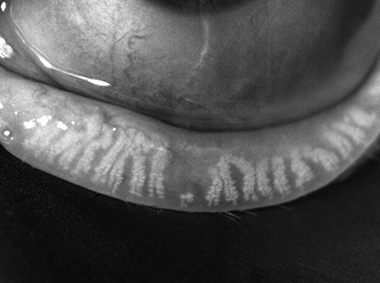
Dynamic Illumination
Surface lighting originates from multiple light sources to minimize reflection.
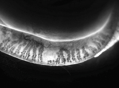
Adaptive Transillumination
Changes to the light intensity across the surface of the illuminator compensate for the lid thickness variations between patients.
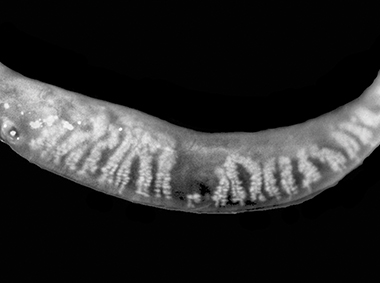
Dual-Mode DMI
A combination of Dynamic Illumination and Adaptive Transillumination offers an enhanced view of meibomian gland structure.
TearScience™ LipiView™ II Reporting1
REAL-TIME VISUALIZATION
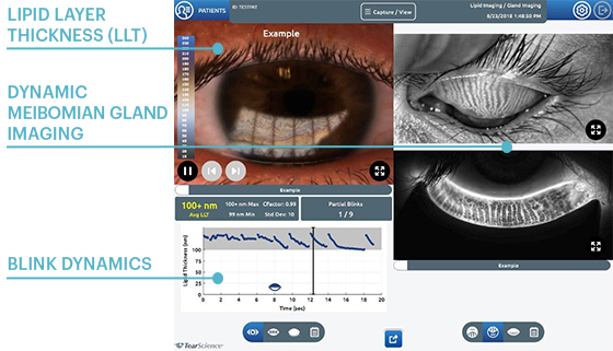
Lipid Layer Thickness (LLT)
Presents lipid layer thickness measurements in an easy to understand color-coded map.
Dynamic Meibomian Gland Imaging
Utilizes advanced illumination technology to capture high-definition images.
Blink Dynamics
Analyzes blink patterns and detects partial blinks. A graphical representation shows fluctuations in lipid layer thickness measurements between each blink.
Visualize and Analyze Blinks1
Analyze Blink Patterns
Computerized analysis of blink patterns objectively quantifies the number of complete and partial blinks. Results are displayed in a frame-by-frame graphical representation that shows fluctuations in lipid thickness measurements between each blink. A video capture allows clinicians and patients to review blinking habits during consultation
Visualize Blink Response
High-resolution video depicts how well ocular lipids disperse in response to a blink through variations in light patterns reflected off the tear film called an interferogram. The system enhances the interference pattern and displays a profile corresponding to an interferometry color scale which has been validated to a known standard for lipid layer thickness measurements with a precision of 1 nm and an accuracy of ±10 nm.
TearScience™ LipiView™ II Features1
- Real-time visualization of the lipid layer to evaluate the dynamic response of lipids to blinking
- Patented noise canceling technology to measure sub-micron thickness of the lipid layer
- Video and analysis of blink dynamics
- High-definition imaging with Dynamic Meibomian Imaging
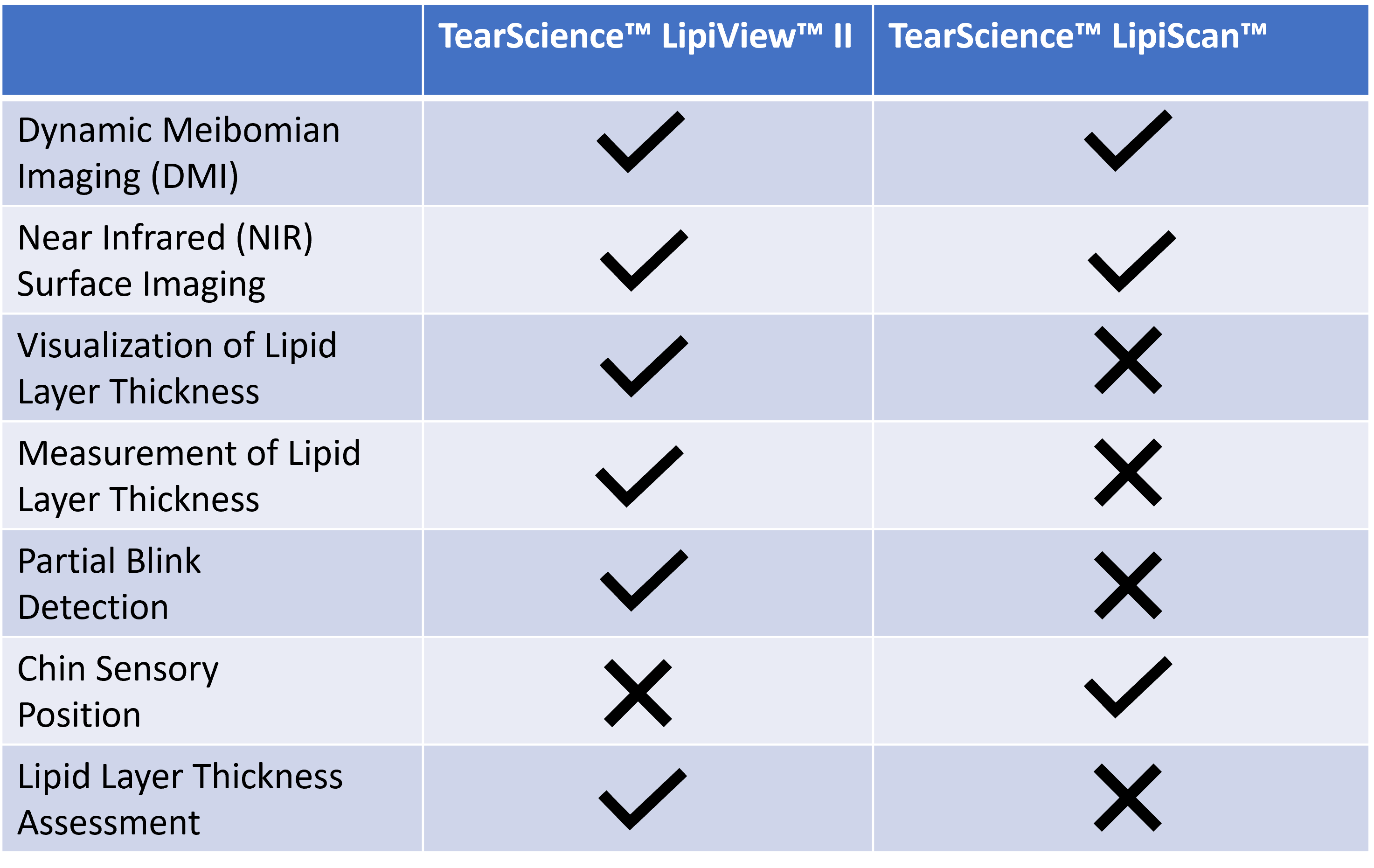
Feature Comparison
REFERENCE
1. TearScience™ LipiView™ II - IFU - 012051-INT, current revision.
© Johnson & Johnson and its affiliates 2024.
For healthcare professionals only. Please reference the Instructions for Use for a complete list of Indications and Important Safety Information and contact our specialists in case of any question.
2024PP12005