CATALYSTM Precision Laser System
CATALYSTM Precision Laser System
")
Instructional Guides
Instructional Guides
Instructional Guides
Clinical Leadership Meets Practice Growth
The CATALYSTM Precision Laser System is specifically engineered to meet your current challenges while priming your practice for the future of cataract surgery. By delivering a purpose-driven combination of outstanding clinical outcomes, immediate practice integration and a premium patient experience, it empowers you to seamlessly elevate your practice today — and seize the opportunities of tomorrow.
The CATALYSTM System is designed for a high standard of clinical precision. It delivers complete capsulotomies and excellent fragmentation so you can give your patients reliably outstanding results.
- Demonstrated high rate of complete 360˚ capsulotomies1
- Multiple centration types — including the scanned capsule for effective positioning2
- Complete softening and segmentation3
- High-quality fragmentation, even in dense cataracts*4
Highly precise, personalized incisions allow you to tailor each procedure to your patients’ unique ocular anatomy for outstanding outcomes
- Wide, side-cut angle (anterior) range of 30 – 150˚ for anterior penetrating and intrastromal incisions5
- Incredible flexibility in incision type and depth for arcuate incisions5
Flexible incision options allow for more personalized and precise surgical procedures.5
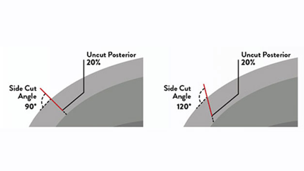
Anterior Penetrating Incisions
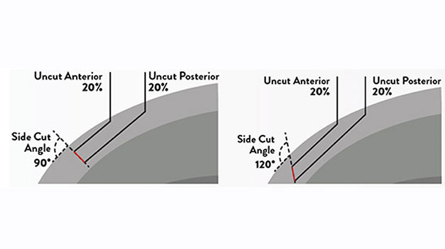
Intrastromal Incisions
The true non-applanating LIQUID OPTICS Patient Interface does not produce corneal endothelial folds, resulting in outstanding incisions.6
The CATALYSTM System’s full-volume, 3D, high-resolution optical coherence tomography (OCT) imaging and INTEGRAL GUIDANCE Technology work together to help you deliver optimal, precise treatment.
- Identifies anterior cornea, posterior cornea, iris, anterior lens and posterior lens to guide laser delivery5
- Refreshes at 0.5 – 2.0 Hz for uninterrupted visualization of the eye throughout treatment5
- Generates accurate pictures of the anterior chamber using 3D OCT imaging data5
Learn more about the CATALYSTM System’s reliably outstanding outcomes (PDF)
The CATALYSTM System can help you elevate your practice by driving growth through increased volume and premium conversion.
- Attract more patients to your practice7
- Convert more patients to premium procedures and IOLs7,8
- Treat more patients7
The CATALYSTM System is designed to fit into your practice with minimal disruption, offering an intuitive and streamlined workflow from start to finish.
- Facilitates quick, single-screen planning with a touchscreen GUI and on-screen keyboard5
- Allows fast, efficient surgical planning and customization5,9
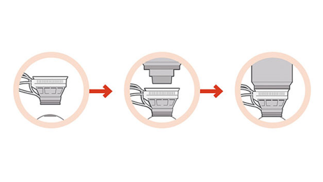
- Guided three-step LOI docking process automatically reduces forces on the eye during treatment5
- Convenience of automated tools with adaptability and control when needed
Learn more about the CATALYSTM System’s ease of integration (PDF)
The CATALYSTM System empowers you to deliver on patient expectations by combining outstanding outcomes and a gentle, streamlined patient experience.
- Quick and gentle docking for patient comfort5
- Personalized procedure from planning to incision5
- Adaptive user interface for outstanding clinical outcomes5
It all starts with an outstanding patient interface.
- True non-applanating design to maintain intraocular pressure6
- Minimizes scleral contact, reducing post-surgery eye redness6
- Offers two patient interface sizes for the ability to dock more patients5
- Not contraindicated for patients with glaucoma5
Learn more about the CATALYSTM System’s patient-focused procedures (PDF)
|
Interface Sizes |
12.0 mm and 14.1 mm |
|
Intraocular Pressure Rise |
10.3 mmHg10 |
|
Imaging Type |
Full-volume, 3D, high-resolution, streaming optical coherence tomography (OCT) imaging |
|
Laser Engine |
Femtosecond diode-pumped, solid state laser engine |
|
User Interface |
Touchscreen, guided user interface with on-screen keyboard |
|
Approximate System Size |
Width: 0.87 m Length: 1.64 m Weight: 340 kg |
FOOTNOTES
*Safety and effectiveness have not been established for cataracts higher than grade 4.
REFERENCES
- 1. Day AC, Gartry DS, et al. Efficacy of anterior capsulotomy creation in FLACS. JCRS. 2014;40(12):2031-2034. REF2014CT0621.
2. Lee YE, Joo CK. Assessment of lens center using optical coherence tomography, magnetic resonance imaging, and photographs of the anterior segment of the eye. Invest Ophthalmol Vis Sci. 2015;56:5512-5518. REF2017CT0142.
3. Conrad- Hengerer I, et al. Effect of femtosecond laser fragmentation of the nucleus with different softening grid sizes on effective phaco time in cataract surgery. JCRS. 2012; 38(11): 1888-94. REF2014CT0024.
4. Dick BH, et al. On the way to zero phaco. JCRS. 2013; 39(9);1442-1444. REF2015CT0248.
5. CATALYSTM cOS7.0 - Op. Manual - 0160-6471, current revision.
6. Talamo J, et al. Optical patient interface in femtosecond laser-assisted cataract surgery. JCRS. 2013; 39(4): 501-510. REF2015CT0427.
7. Ondrias D, Integrating laser cataract surgery. Optometry Times. March 2015. REF2015CT0235.
8. Aker A, Femto-cataract: Why the business model works. Ophthalmology Times, May 2015;40(8):1, 20-21. REF2015CT0232.
9. Friedman NJ. A comparative look at femtosecond lasers for cataract surgery, Part 1. OphthalmologyWeb, 2014. REF2015CT0079.
10. Schultz T, et al. Intraocular pressure variation during femtosecond laser-assisted cataract surgery using a fluid-filled interface. JCRS. 2013; 39:22-27. REF2014OTH0027.
PP2022CT4108 v2